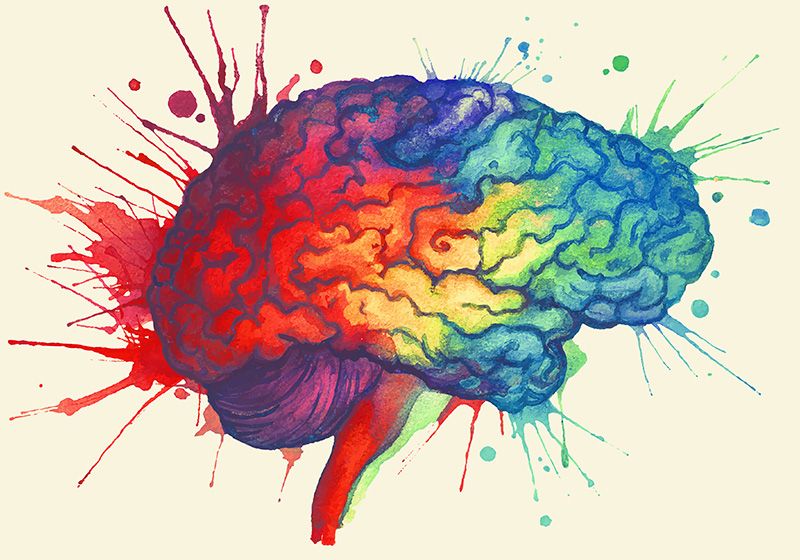
Using transcriptomics and machine learning, scientists observed the formation of new neurons in the adult human brain.
Various tissues of the human body—the skin, the mucosal lining of the digestive tract, and blood—are regularly rejuvenated through the formation of new cells, a process crucial to their proper functioning.1 Similarly, certain brain functions, such as memory formation and mood regulation, involve the birth of new neurons.2 Though studies reported neurogenesis in adult rodent brains, contradictory evidence has fueled a decades-long controversy on whether and how new neurons form in the adult human brain. In a new study published in Science, researchers at Karolinska Institute used transcriptomics and machine learning to show that neural progenitor cells and their successors, newly born immature neurons, are present from childhood to old age in humans.3
Jonas Frisén is a stem cell biologist at Karolinska Institute who studies the role of cell replacement in physiological conditions.
Stefan Zimmerman
“This gives us an important piece of the puzzle in understanding how the human brain works and changes during life,” said Jonas Frisén, a stem cell biologist at Karolinska Institute and author of the study, in a statement. “Our research may also have implications for the development of regenerative treatments that stimulate neurogenesis in neurodegenerative and psychiatric disorders.”
In the early 1900s, scientists believed that the generation of new neurons stopped soon after birth. Along came a barrage of experiments in the late 20th century that sowed seeds of doubt regarding the theory. Using techniques like genetic tracing, DNA labeling, and live imaging, researchers began peering into the brains of rodent models, focusing on the hippocampus, a region crucial for memory, learning, and mood control.2 They observed dividing progenitor cells and new immature neurons in animals of varied ages. Emboldened with these results, the field set its sights on the human hippocampus, but that’s when things fell apart. Technical challenges like poor translation of neural progenitor markers from rodent studies to human samples and sparse numbers of the progenitors among other neurons created inconsistencies in the observations from different groups.4,5
To overcome these barriers, Frisén and his team analyzed the RNA signatures of cells from childhood to old age using single nucleus RNA sequencing. First, they isolated more than 100,000 nuclei from postmortem hippocampal tissue of humans from zero to five years of age (“childhood cohort”), sequenced the RNA, and looked for markers associated with neural progenitor cells. Consistent with previous findings by Frisén and other groups, they identified neural progenitors and immature neurons in these samples. Similarly, they collected more than 200,000 hippocampal nuclei from individuals of 13 to 78 years of age (“adult cohort”) and analyzed their RNA expression profile. To reliably detect neural progenitors in the adult cohort, the team trained a machine learning model on the childhood cohort data to identify the signatures of these cells. The model reported the presence of neuronal progenitors and immature neurons in adult hippocampal tissue, whose RNA signatures matched early neuronal development stages.
“We have now been able to identify these cells of origin, which confirms that there is an ongoing formation of neurons in the hippocampus of the adult brain,” Frisén said.
The researchers also noted interindividual variation across ages. Two out of the 14 adults had significantly higher number of progenitors, while five did not show evidence of continued neurogenesis. However, the study authors have yet to investigate if these differences arose from technical variability or underlying biological mechanisms.
Since many neurological diseases affect the viability of brain neurons, the team believes that their findings could spark the investigation of the effects of pathology and genetics on the lifelong birth of neurons.


